
Cerebrovascular reserve capacity describes how well the brain's vessels can adapt to changing conditions. It is reduced in advanced atherosclerosis of the large brain arteries and is a risk factor for ischaemic stroke.
In the study led by Prof. Ilko Maier, 25 healthy volunteers were examined during a standardised hyperventilation–apnoea test with both neurovascular ultrasound and real-time phase-contrast MRI, in the middle and posterior cerebral arteries. Both methods reliably showed the expected changes in blood flow velocity; absolute values were somewhat lower with MRI.
The authors consider real-time MRI a promising technique for assessing reserve capacity in various intracranial vessels.
Original article: Horstmann C, Hofer S, Dechent P, Bähr M, Maier I. Real-time phase-contrast MRI can be used to quantify cerebrovascular reserve capacity – a comparative study to neurovascular ultrasound in healthy subjects. Neuroimage Rep 2026;6(1):100332. doi:10.1016/j.ynirp.2026.100332 (PMID 41993702)
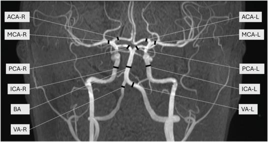
Cover image (from the publication above): Fig. 1. TOF-MRA of the intracranial arteries with marked locations for intracranial flow measurements with the RT-PC MRI. Measurement locations are marked as left (L) or right (R) site. MCA: medial cerebral artery; ACA: anterior cerebral artery; PCA: posterior cerebral artery; BA: basilar artery; VA: vertebral artery; ICA: intracranial carotid artery.